Lasing as a tool for detection of protein aggregates
2021-10-06
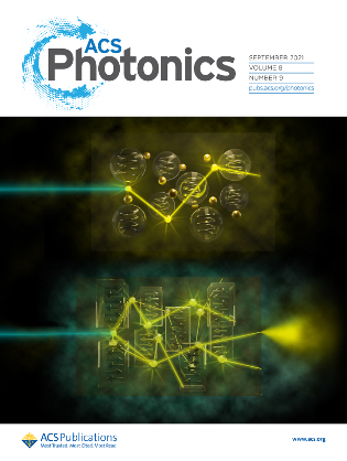
Scientists from the Laboratory of Ultrafast Processes at the Faculty of Physics of the University of Warsaw have developed a method that allows to detect small protein aggregates (oligomers), the presence of which in the nervous tissue and the cerebrospinal fluid may signal the early stages of neurodegenerative diseases such as the Alzheimer's or Parkinson's diseases. The method is based on the use of the phenomenon of stimulated emission to amplify the light emitted spontaneously by dye molecules associated with protein aggregates. A paper describing this research is promoted on the main cover of the September issue of ACS Photonics.
The development of neurodegenerative diseases such as the Alzheimer's or Parkinson's diseases is associated with the accumulation of aggregates of misfolded proteins, the so-called amyloids. The presence of large amyloid deposits in the patient's brain is characteristic of, for example, the Alzheimer's disease. The current state of knowledge on the development of neurodegenerative diseases indicates, however, that the formation of large deposits is only the last stage of the disease, and its development may begin even decades before the appearance of clinical symptoms. In the early stages of the disease, small amyloid aggregates (oligomers) are formed, the detection of which could allow the diagnosis of the Alzheimer's disease many years earlier than it is currently possible.
Unfortunately, current diagnostic techniques are insensitive to the presence of oligomers. The fluorescent marker commonly used for the detection of amyloids, thioflavin T, detects amyloid aggregates only when they have reached the stage of elongated fibrils. In the presence of smaller oligomers, thioflavin does not emit fluorescence of detectable intensity upon optical excitation. However, the work published in ACS Photonics shows that the emission properties of thioflavin T can be used, as long as the sample stained with this dye is excited by a laser pulse so strong that the sample contains more excited molecules than molecules in the ground state (this is the so-called population inversion, also crucial for the operation of lasers). Under such conditions, a photon emitted spontaneously by an excited thioflavin molecule may with a high probability interact with another molecule in the excited state and lead to the emission of a second identical photon as a result of the stimulated emission process. Subsequent acts of stimulated emission lead to an avalanche increase in the number of photons and the generation of the so-called amplified spontaneous emission (ASE), similar to the light emitted by a laser. Since the properties of the light emitted by this phenomenon depend on the path traveled by photons in the sample, and this path is related to the sample structure, the structural properties of the sample (in particular the presence of protein oligomers) can be characterized by ASE measurements.
For more information, please contact Piotr Hańczyc or Piotr Fita from the Laboratory of Ultrafast Processes.
In the Laboratory of Ultrafast Processes, students can take a paid internship involving research on optical studies of protein aggregation. Interested students should contact Piotr Hańczyc.
RELATED PUBLICATIONS:
- P. Hanczyc, P. Fita, Laser Emission of Thioflavin T Uncovers Protein Aggregation in Amyloid Nucleation Phase, ACS Photonics, 8 2598-2609 (2021)
- P. Hanczyc, M. Procyk, C. Radzewicz, P. Fita, Two-photon excited lasing of Coumarin 307 for lysozyme amyloid fibrils detection. J. Biophotonics 2019, 12, e201900052


